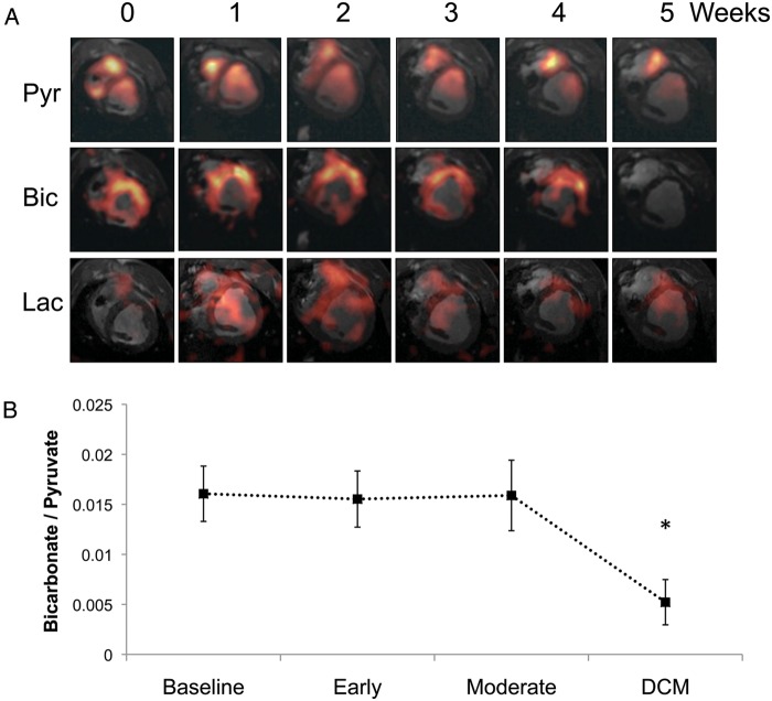
Figure 4.
Hyperpolarized 13C magnetic resonance imaging (MRI) results describing alterations to pyruvate dehydrogenase complex (PDC) flux and [13C]lactate production with the pathogenesis of dilated cardiomyopathy (DCM), following infusion of hyperpolarized [1-13C]pyruvate. (A) Representative pyruvate (Pyr, top), bicarbonate (Bic, middle), and lactate (Lac, bottom) 13C MR images taken from the same pig and at weekly intervals during the pacing protocol, until DCM developed. The images displayed for each metabolite were selected from the same, mid-papillary slice and in the same respiratory cycle. Signal intensity in the pyruvate image was scaled based on 15–100% of the maximum pyruvate signal at week 0, whereas the bicarbonate and lactate signal intensities were scaled based on 15–100% of the maximum bicarbonate signal intensity at week 0. (B) Relative changes to PDC flux with DCM in five pigs.

