
Figure 6.
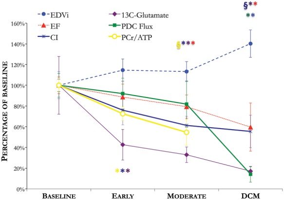
An overview of LV metabolic, energetic, structural, and functional remodelling, measured non-invasively using magnetic resonance imaging (MRI) and magnetic resonance spectroscopy (MRS), throughout the development of tachycardia-induced dilated cardiomyopathy (DCM). *P < 0.05 compared with baseline; §P < 0.05 compared with the early time point. CI, cardiac index; EDVi, end-diastolic volume index; PCr, phosphocreatine; PDC, pyruvate dehydrogenase complex.