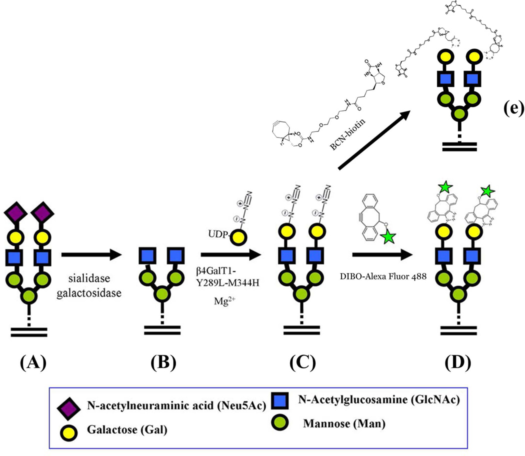
Figure 3.
Schematic diagram showing the GlcNAc specific transfer of GalNAz and its conjugation with Alexa Fluor 488 fluoroprobe. A: On the HeLa cell surface, the GlcNAc moiety in glycoconjugates is galactosylated and sialylated. A representative cell surface glycan is shown as a biantennary N-glycan that is in fully glycosylated form. B: When the cells are treated with the α1-3,6,8 sialidase and β1-4-galactosidase, the terminal sialic acid and β1-4-linked galactose are sequentially removed from the glycans, thereby exposing the GlcNAc moiety at the nonreducing end. C: The pretreated cells are incubated with the Y289L-M344H-β4Gal-T1 enzyme in the presence of UDP-GalNAz and Mg2+, transferring GalNAz to the non-reducing end of the GlcNAc moiety. D: When the glycan modified cells are treated with DIBO Alexa Fluor 488, the fluoroprobe molecules selectively conjugate with the GalNAz moieties, forming a covalent bond with them. Now the cells are site-specifically labeled; the fluorescence detected on the cell surface corresponds to the amount of the free GlcNAc moiety present on them. E: Copper-free click reaction with BCN-biotin to detect free GlcNAc residues by Western blot analysis or streptavidin-magnetic bead isolation.

