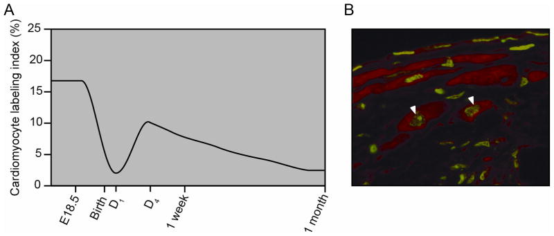
Figure 2.

The neonatal and adult mammalian hearts are capable of cellular proliferation. A) Relative cardiomyocyte proliferation from BrdU labeling and tritiated thymidine incorporation studies of neonatal and adolescent mice support the notion that the heart’s proliferative capacity decreases with age (see references #34 and #35). B) BrdU labeling studies following injury reveal proliferation of murine cardiomyocytes in the border region of the adult mouse heart. Arrowheads mark BrdU labeled cardiomyocyte nuclei, which are green and are immunostained with α-actinin, which is red. Adapted from Naseem et al. Physiol Genomics, 2007.38 Am Physiol Soc, used with permission.
