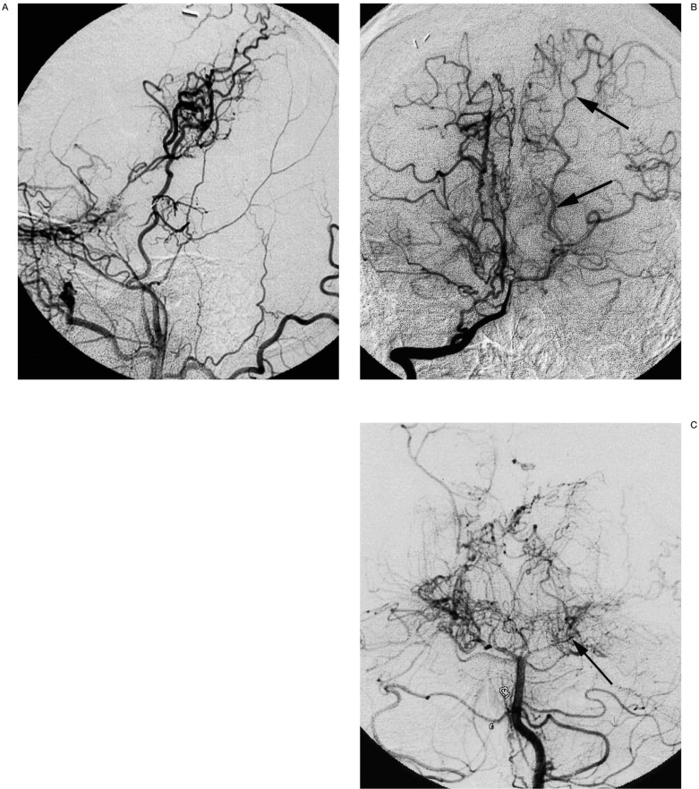
Figure 2.
A 23-year-old man with moyamoya disease whose initial ischaemic events occurred 13 years ago. Left common carotid artery injection (A, lateral view) shows occlusion of internal carotid artery below the origin of ophthalmic artery. Good collateral though the surgical anastomosis is observed. Right vertebral artery injection (B, frontal view) shows the patent left posterior cerebral artery (arrows). Five months after this angiogram, the patient developed right homonymous hemianopsia due to occlusion of the P2 portion of left posterior cerebral artery (C, frontal view, arrow).

