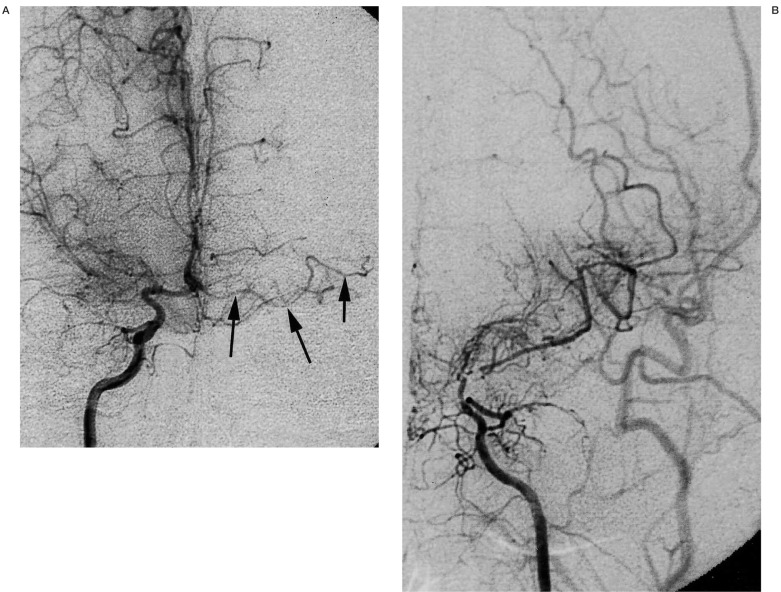
Figure 3.
A 30-year-old man with moyamoya disease who developed transient ischaemic attack 15 years ago. Right internal carotid injection (A, frontal view) shows occlusion of right middle cerebral artery and minimal development of moyamoya vessels. Arrows indicate left accessory middle cerebral artery without any stenotic changes. Left internal carotid injection (B, frontal view) shows marked stenosis at the terminal portion of internal carotid artery.

