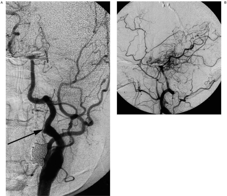
Figure 4.
A 54-year-old woman with moyamoya disease who initially developed ischaemia 10 years ago and subsequently developed subarachnoid haemorrhage 5 years ago due to ruptured basilar bifurcation aneurysm. Left common carotid injection (A, frontal view;B, lateral view) shows the previously patent, but recently occluded left internal carotid artery and persistent primitive hypoglossal artery (arrow) with development of moyamoya vessels. Basilar bifurcation aneurysm is surgically clipped.

