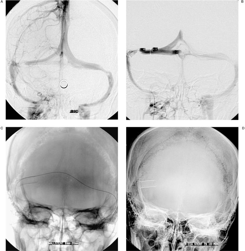
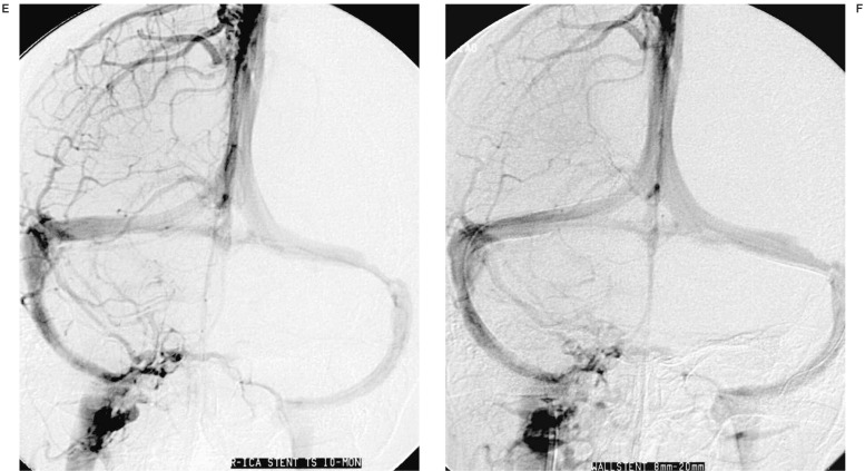
Figure 1.
Lateral venous phase and venous angiography showed bilateral transverse sinus stenosis (A,B), a 9 F guiding catheter was navigated with a 0.018 in microwire to the proximal right jugular bulb, the microwire was passed through the stenosed transverse sinus and navigated to the opposite jugular bulb, a self-expanded Wallsten stent of 8 × 20 mm was deployed at the stenosis site of right transverse sinus (C), the stenosis segment was completely dilated (D).Ten months angiographic follow-up showed the stent located at the original deployed site and the stented sinus kept good patency (E,F).


