
Figure 3.
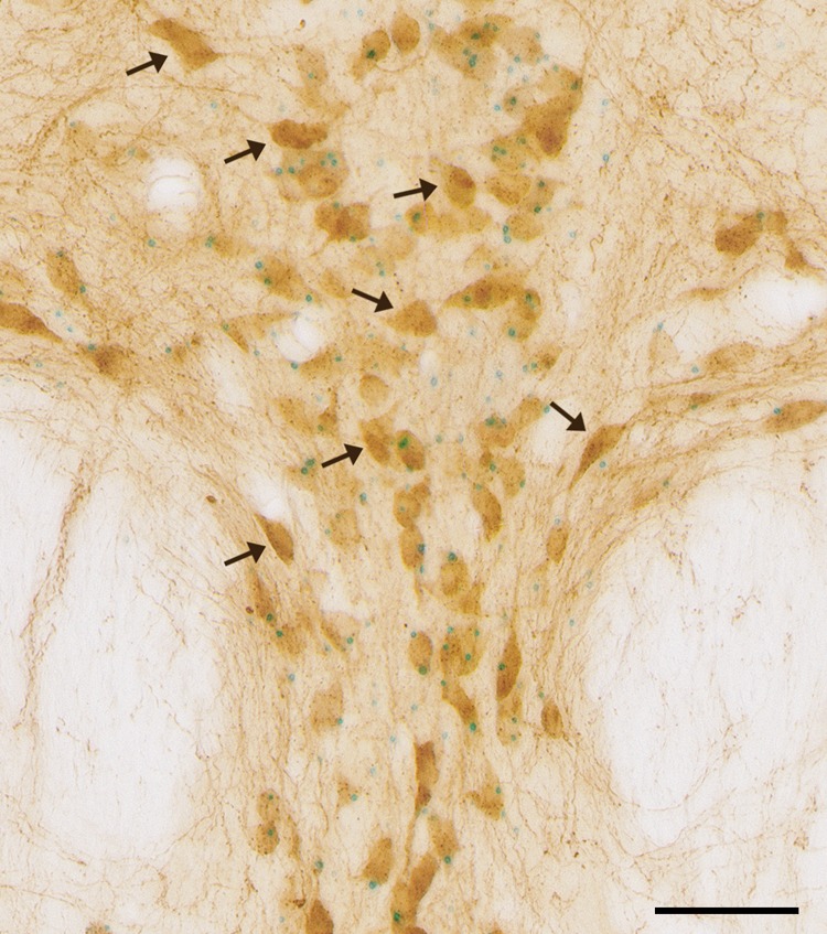
High magnification image depicting the existence of 5-HT1A-negative neurons in the dorsal raphe of 5-HT1A-iCre/R26R mice. 5-HT1A receptor mRNA expression (blue dots) and 5-HT-IR (brown) were detected using combined X-Gal staining and immunohistochemistry in 5-HT1A-iCre/R26R mice. The black arrows points to 5-HT-IR single-labeled neurons. Images were taken at 40× magnification (scale bar 50 μm).