
Figure 6.
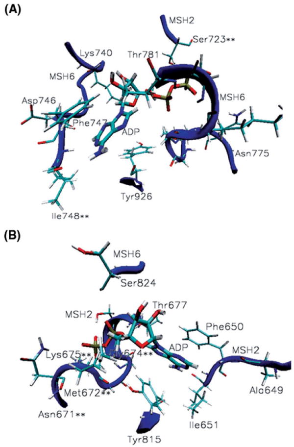
MutSα nucleotide binding sites. (A) ADP binding contacts at the ATPase domain of MSH6: adenine stacks between Phe747 and Tyr926 and makes specific contacts with Lys740, Asn746-Ile748; the phosphate group binds at the P-loop Asn775-Thr781 and hydrogen bonds Ser723 from the alternate binding site in the mismatched complex only. (B) ADP binding contacts at the ATPase domain of MSH2: adenine stacks between Phe650 and Tyr815 and makes specific contacts with Ala649-Ile651; the phosphate group binds at the P-loop Asn671-Thr677 and hydrogen bonds Ser824 from the alternate binding site. Noteworthy is the significant presence of cancer causing mutations among the ADP binding contacts of both subunits (residues marked with **), 25% of them.