Figure 5.
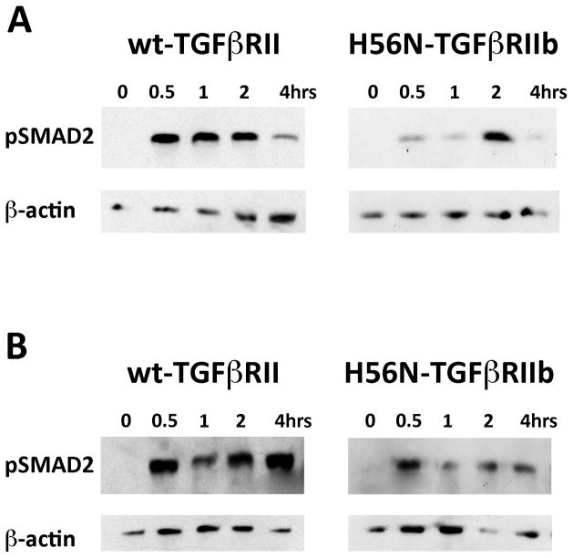
TGFβ1 and TGFβ2 signaling in dermal fibroblasts. A–B. Cultured fibroblasts were isolated from normal patient with wt-TGFbRII and ANV I-1 patient with H56N-TGFbRIIb mutation. Representative Western blot analyses shown for pSMAD2 protein expression in wt-TGFbRII or H56N-TGFbRIIb fibroblasts stimulated with either TGFb1 (A) or TGFb2 (B) for indicated time points. Corresponding b-actin blots shown. (A) H56N-TGFbRIIb dermal fibroblasts exhibit delayed TGFβ1 signaling compared to normal dermal fibroblasts. (B) H56N-TGFbRIIb dermal fibroblasts exhibit decreased TGFβ2 signaling compared to normal dermal fibroblasts.

