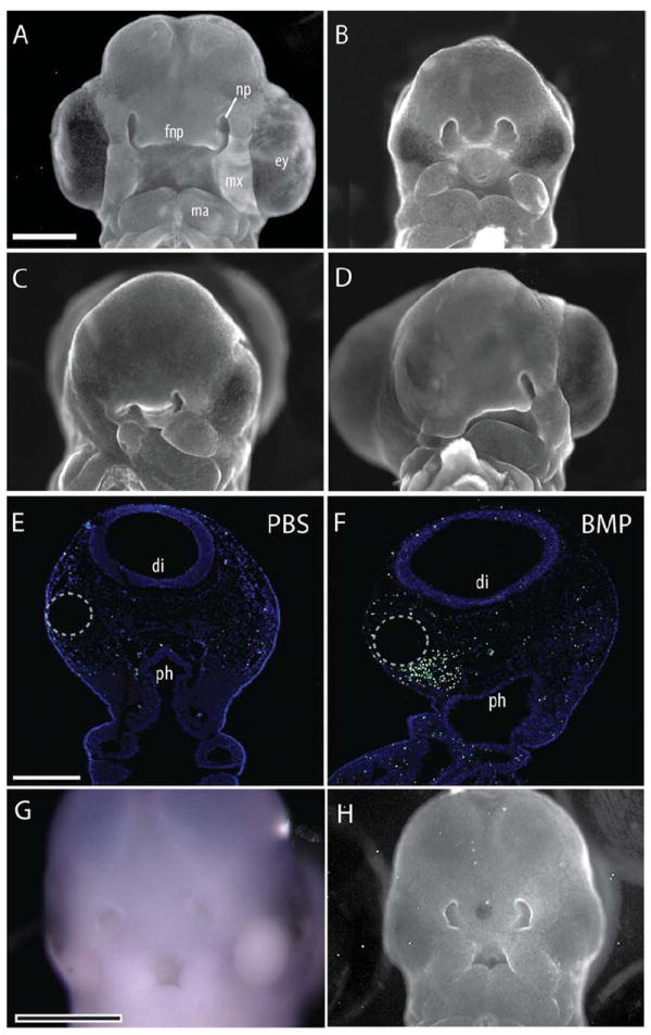
Fig. 1.

Facial defects were evident 72 hr after exogenous BMP activation. A: A normal embryo illustrating facial morphology at HH22. B: Injection of hybridoma cells expressing anti-SHH antibodies (5E1) into the neural tube blocked SHH signaling within the forebrain and produced facial malformations. Sham rescue using a PBS-soaked bead did not alter morphology (n=10). Embryos exhibited microcephaly, microphthalmia, and narrowing of the FNP. The maxillary and mandibular processes were also medially rotated and smaller than normal. C: When a BMP-4-soaked bead was placed into the into the head mesenchyme of 5E1-treated embryos, additional severe defects of the eye, face, and jaw were observed. The side of the BMP bead placement was more severely affected (n=13). D: Activation of the BMP pathway without blockade of SHH signaling also produced severe facial malformations (n=8), with the side of bead placement more severely affected. E: TUNEL staining of coronal sections of the head of control embryos illustrates minimal cell death. F: In contrast, a dramatic increase in cell death was evident at 24 hr after treatment with BMP-4-soaked beads (25 μg/mL) (n=11). Cell death was most concentrated in the mesenchyme surrounding the bead (dotted circle). G: Whole mount in situ hybridization for Shh illustrates that activation of the BMP pathway did not restore Shh expression (n=25). H: When BMP-soaked beads were placed into the FNP of 5E1-treated embryos at HH18, morphology was not restored at HH22 (n=8). Scale bar in A–D, G, H = 1 mm; in E,F = 300 μm. fnp, frontonasal process; np, nasal pit, ey, eye; mx, maxilla; ma, mandible; di, diencepalon; ph, pharynx.
