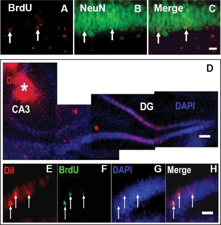
Fig. 1.
Double immunofluorescent staining for BrdU (red, A) and NeuN (green, B) to identify newborn neurons (yellow after merge, C) in the dentate gyrus of hippocampus from rats examined 35 days after TBI. Micrographs (D) show location of DiI injection in the CA3 region (indicated by white asterisk). In the CA3 region, axons projected from granule neurons in the dentate gyrus will take up injected DiI to their cell bodies. Co-localization (merge, H) of BrdU-positive nuclei (green, F) within retrogradely DiI labeled (red, E) granule cells were examined at 35 days after TBI. Scale bar = 25 μm (C, H). Scale bar = 50 μm (D).

