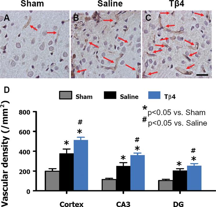
Fig. 2.
Delayed Tβ4 treatment increases vascular density in the injured cortex, ipsilateral dentate gyrus, and CA3 region 35 days after TBI. Arrows show vWF-stained vascular structure. TBI alone (B) significantly increases the vascular density in the injured cortex compared to sham controls (A, P < 0.05). Tβ4 treatment (C) further enhances angiogenesis after TBI compared to the saline-treated groups (P < 0.05). The density of vWF-stained vasculature in different regions is shown in (D). Scale bar = 25 μm (C). Data represent mean + SD. *P < 0.05 vs Sham group. #P < 0.05 vs Saline group. N (rats/group) = 6 (Sham); 9 (Saline); and 10 (Tβ4).

