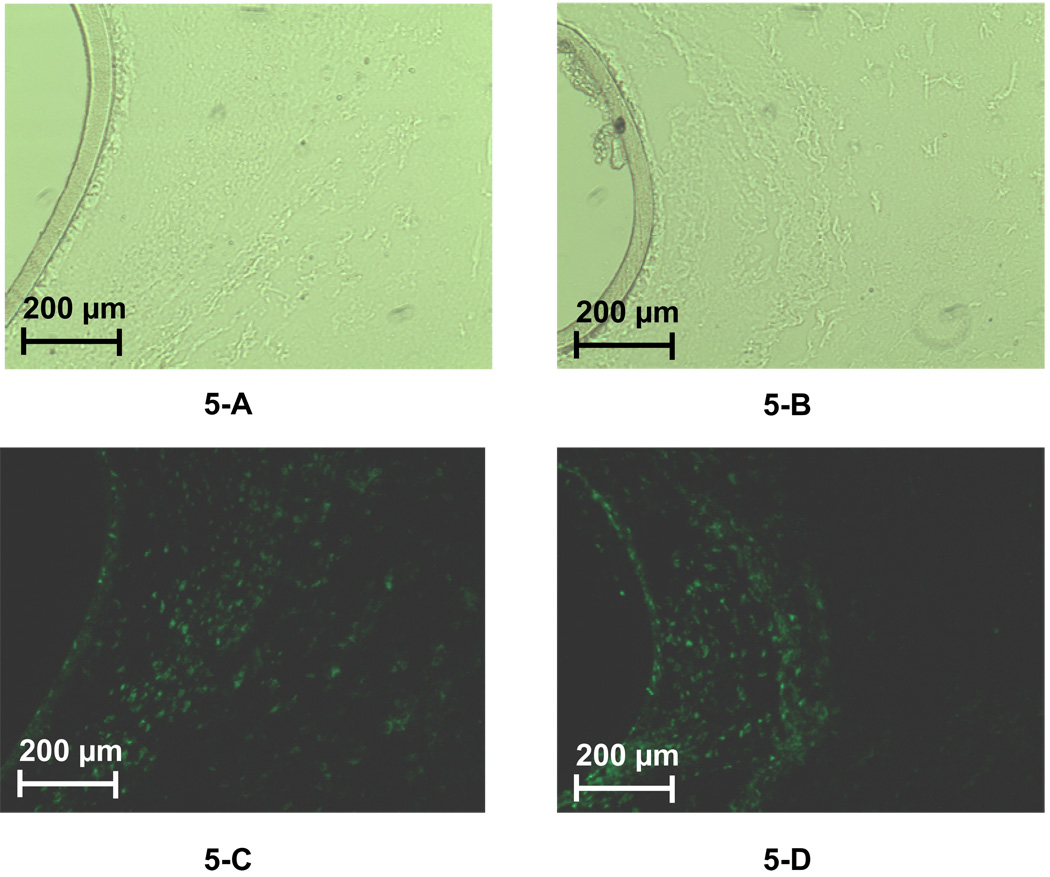
Figure 5.
Immunofluorescence of labeled macrophages in the tissue space surrounding the membrane of 5-day (5-A and 5-C) and 7-day (5-B and 5-D) implanted microdialysis probes (PC membrane). A 20× objective was used for all the images. For each fluorescence image, there is a paired image taken under normal light with the same magnification to reveal the location of the probe membrane. Figure 5-A and 5-B were obtained using a normal microscope lamp. Figures 5-C and 5-D were obtained using the fluorescence lamp with a filter.

