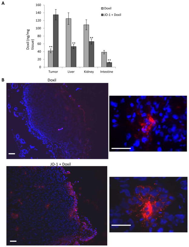
Figure 3. Liposomal doxorubicin/Doxil biodistribution.
Mice carrying HCC1954 tumors were intravenously injected with PBS, Doxil (1 mg/kg) or, JO-1 (2 mg/kg) followed by Doxil one hour later. Two hours after PBS or Doxil injection, mice were sacrificed, blood was flushed from the system, and organs were collected to analyze the presence of Doxil in tissues using antibodies against PEG. A) Tissue lysates were subjected to ELISA for JO-1. n=3. The differences between the two groups are significant (**P<0.01). B) Immunfluorescence analysis of tumor sections with anti-PEG antibodies. The scale bar is 20 μm. Notably, free PEG is poorly detected by ELISA or immunohistochemistry (13). Effective binding is greatly enhanced in PEG conjugates such as Doxil.

