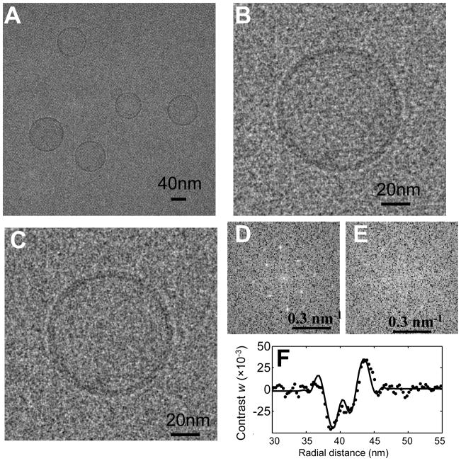
Fig. 5.
Effect of the crystal lattice removal process on the images of DPhPC liposomes tethered on the 2D crystal. (A) Cryo-EM image of liposomes tethered to a 2D crystal. (B–C) Cryo-EM image of an individual liposome before and after the removal of crystal information. The images were Gaussian-filtered (half power at 2.0 nm) for display. (D–E) The center quarter of the power spectrum of the image before and after masking the spots. (F) Circularly averaged experimental liposome image contrast w (dots) compared with model (line). The image was taken on film, without energy filtering, using the Tecnai F20 electron microscope at 200 keV. The defocus was 2.20 μm and the B factor was estimated to be 1.86 nm2.

