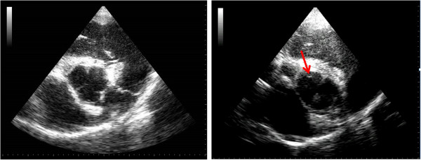
Figure 3.

Right parasternal short axis echocardiograms of the aortic valve (AoV). The image on the left shows a view of a normal AoV in the centre of the image as well as the landmarks described in the optimisation study. The image on the right is from aortic valve prolapse (AVP) of the non-coronary cusp (NCC). The images shows the edges of the right and left coronary cusps and the red arrow is highlighting the NCC not being present within the imaging plane as the other two are.
