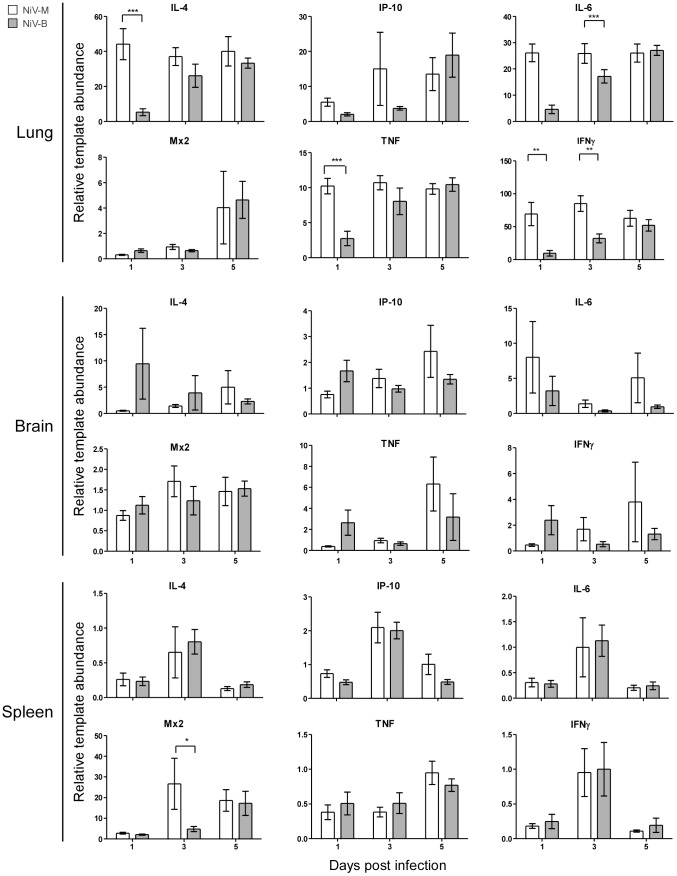
Figure 4. Host gene expression in lung, brain and spleen tissue of hamsters is differentially regulated during infection with Nipah viruses.
Quantitative RT-PCR for IL-4, IP-10, IL-6, Mx2, TNF and IFNγ was performed on lung, brain and spleen tissues from groups of 9 hamsters inoculated with 105 TCID50 of NiV-M (white bars) or NiV-B (gray bars) at the indicated time points. Data are shown as the fold-change of each gene over uninfected controls and normalized to an internal reference gene (RPL18). Error bars represent the SEM. A 2-way ANOVA with Bonferroni's post-test was used to determine statistical significance between viruses (* = p<0.05, ** = p<0.01 and *** = p<0.001).

