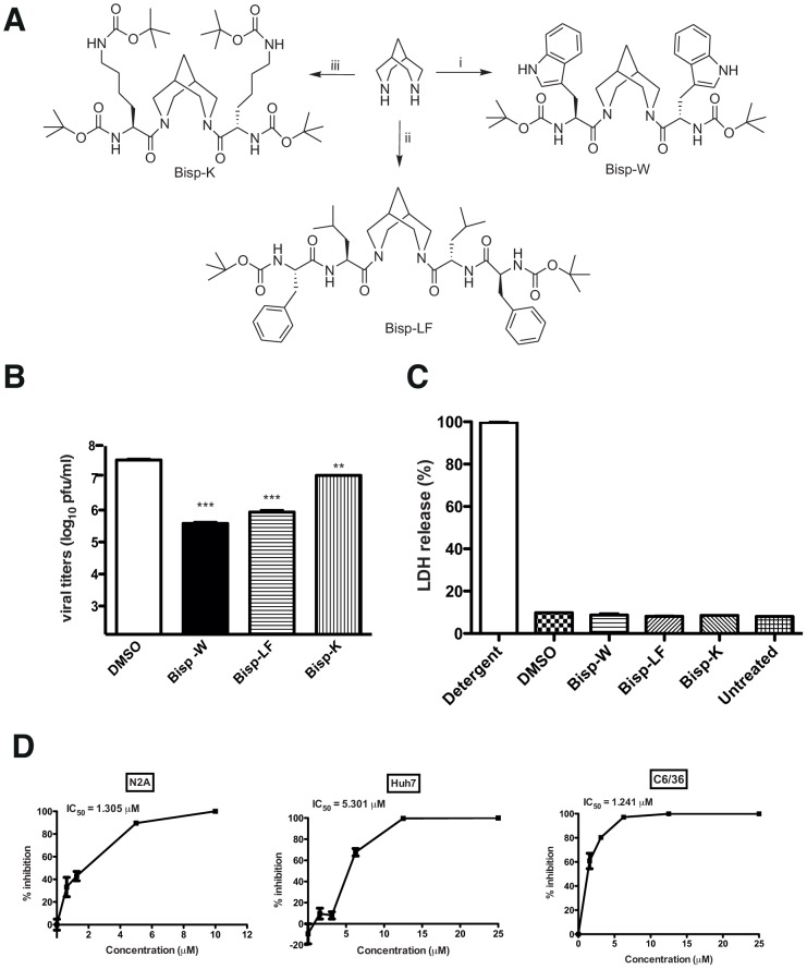
Figure 3. Synthesis of amino acid conjugates of bispidine.
(A) Structure of bispidine conjugated with tryptophan (Bisp-W), lecine+phenylalanine (Bisp-LF) and lysine (Bisp-K). (B) Viral titers were determined by plaque assay of N2A cell culture supernatants (22 h pi) infected with JEV and treated with 5 µM of derivatives of bispidine. *** P = 0.0004 and 0.0004, ** P = 0.0034 and as determined by two-tailed, t-test. Error bars represent Mean ± SEM of three replicates. (C) Cytotoxicity was measured by lactate dehydrogenase (LDH) assay from culture supernatants treated with 5 µM of the indicated bispidine conjugates or DMSO. LDH released from cells incubated with detergent buffer was used as 100% LDH release. (D) IC50 value for Bisp-W in the indicated cell lines was estimated by measuring viral titers in cell culture supernatants (22 h pi) infected with JEV and treated with the indicated concentration of Bisp-W. Error bars represent Mean ± SEM of three replicates. All the data are representative of experiments performed at least twice with three replicates.

