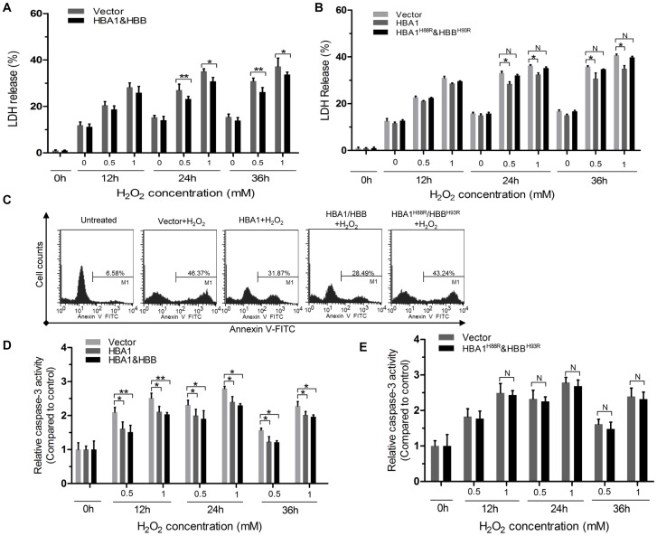
Figure 6. Either HBA1/HBB or HBA1, but not HBA1H88R/HBBH93R overexpression protects cells against oxidative stress-induced damage.
(A) The viability of SiHa cells treated with 0, 0.5, and 1 mM H2O2 for 12, 24 and 36 h as extracellular oxidative stress was evaluated by the LDH release assay. SiHa cells transfected with the HBA1/HBB-expressing vectors (black column) showed increased resistance against H2O2-induced cell compared to cells transfected with the control vector (gray column). Similar experiments were preformed in both HBA1-overexpression cells and HBA1H88R/HBBH93R-overexpression cells (B). SiHa cells were transfected with the indicated plasmids. Forty-eight hours later, cells were treated with 1 mM H2O2 or left untreated for 22 h. The percentage of apoptotic cells was monitored by Annexin V staining followed by FACS analysis (C). SiHa cells were transfected with the indicated plasmids and forty hour later, were treated as in (A), Caspase-3 activity in different group was measured using specific caspase substrate AcDEVD-pNA as described under MATERIALs AND METHODs. Values of the absorption were expressed as control which was set as 1 (D and E). All experiments were performed in triplicate and were repeated at least three times, and the results are expressed as means ± S.E.M.*P<0.05; **P<0.01; N P>0.05.

