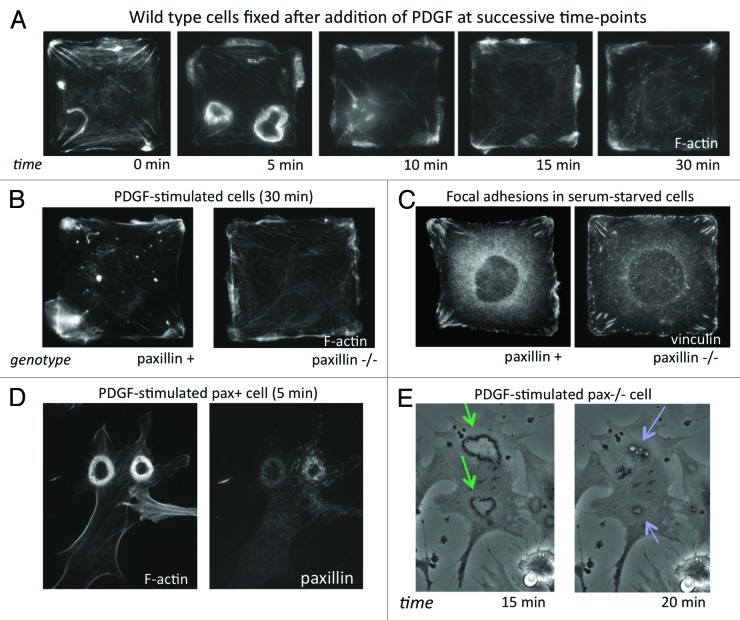
Figure 2. (A) Time-course of the motile response to PDGF (25 ng/ml) stimulation in wild type (pax+) mouse embryonic fibroblasts (MEFs). Cells were fixed without (0 min) and at 5, 10, 15 and 30 min after addition of PDGF and stained with Alexa-488 phalloidin to label F-actin. (B) Pax+ and pax−/− MEFs stimulated with PDGF for 30 min, fixed and stained with Alexa-488 phalloidin. (C) Vinculin labeling by immunofluorescence in serum-starved pax+ and pax−/− cells on square islands showing differences in FA distribution. (D) Pax+ MEF fixed 5 min after addition of PDGF and stained for F-actin and paxillin showing large circular dorsal ruffles (CDRs). (E) Phase-contrast time-lapse video of live pax−/− MEFs stimulated with PDGF (20X). Formation of CDRs (green arrows) persisted for more than 15 min and protrusions were internalized into macropinocytic vesicles (blue arrows) by about 20 min. In pax+ cells, CDR formation and internalization was complete by 10 min after addition of PDGF.

An official website of the United States government
Here's how you know
Official websites use .gov
A
.gov website belongs to an official
government organization in the United States.
Secure .gov websites use HTTPS
A lock (
) or https:// means you've safely
connected to the .gov website. Share sensitive
information only on official, secure websites.
