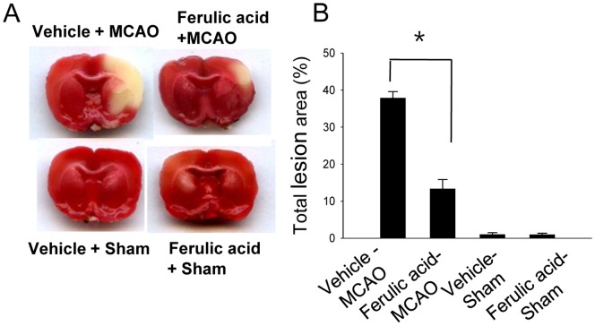
Figure 1. The neuroprotective effect of ferulic acid in focal cerebral ischemia.
Representative photos of TTC stain in the cerebral cortices of vehicle+sham, ferulic acid+sham, vehicle+middle cerebral artery occlusion (MCAO), ferulic acid+MCAO animals. Animals were treated with vehicle or ferulic acid prior to MCAO. Brain sections were stained by TTC (A). The ischemic area remained white, while the intact area was stained red. The percentage of ischemic lesion area was calculated by the ratio of the infarction area to the whole slice area (B). Data are means ± S.E.M. * P<0.05.

