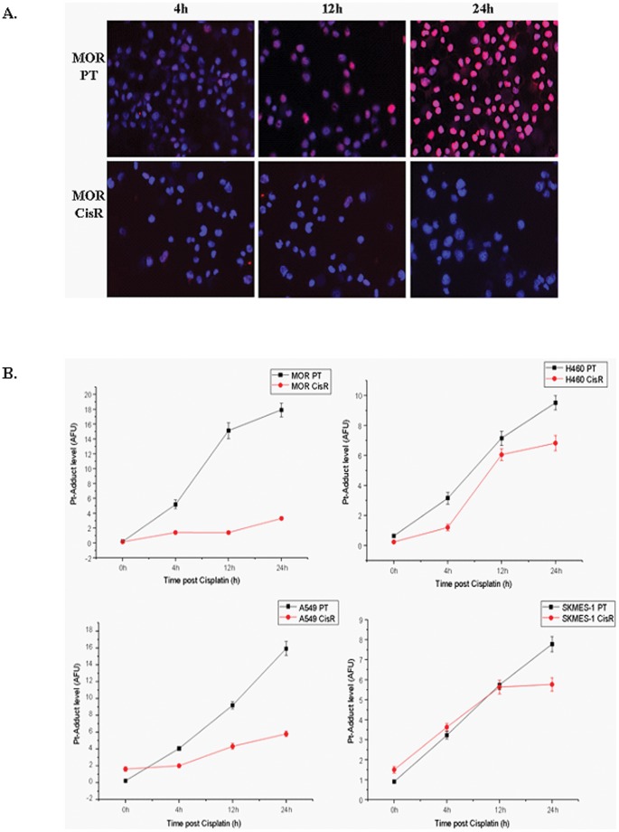
Figure 11. Cisplatin-DNA adduct formation and immunofluorescence.
Lung cancer cell lines were treated with cisplatin for up to 24 h and fixed on Superfrost Gold Slides using ice-cold methanol. Cells were stained overnight at 4°C using a primary antibody that specifically recognizes CDDP-GpG DNA adducts (RC-18). Antibody binding was detected using an anti-rat Cy3®-labelled antibody and counterstained using DAPI (1 µg/ml (w/v). Images were acquired on an Axioplan fluorescence microscope (A). Adducts were quantified and measured as arbitrary fluorescence units (AFU’s) upon normalisation of integrated antibody-derived fluorescence from 200 individual nuclei of the corresponding DNA content. Data are presented as the mean AFU ±95% confidence interval (CI) from three independent experiments (B).

