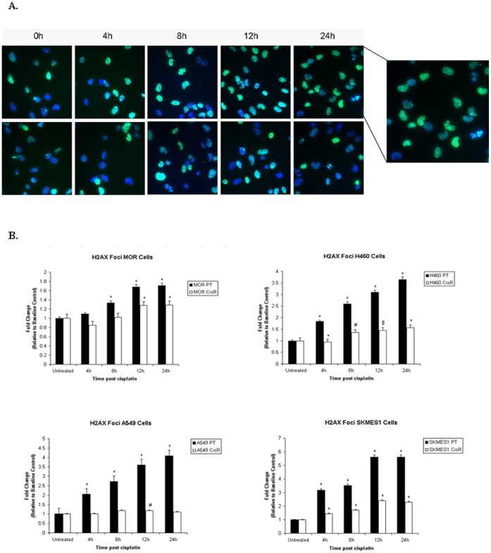
Figure 12. Measurement of γH2AX foci formation and DNA damage.
Following treatment of parent and chemoresistant cells with cisplatin for 4, 8, 12 and 24 h, cells were fixed in formaldehyde and incubated with a primary rabbit anti-human anti-phospho-histone 2AX (Ser139) antibody. Cells were subsequently labelled with an Alexafluor 488-labelled goat anti-rabbit secondary antibody and Hoechst 33342 nuclear stain prior to analysis by high content analysis using the InCell Analyser 1000 (A). Data are expressed as Mean ± SEM from three independent experiments (n = 3) (#p<0.05, $p<0.01, *p<0.001) (B).

