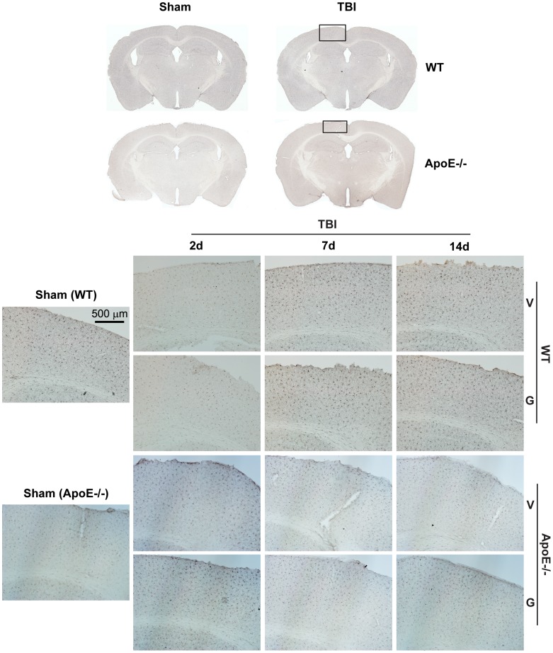
Figure 7. Microglia are not significantly activated by mrTBI in either WT or apoE−/− mice.
Microglial activation in sham and injured WT and apoE−/− mice was assessed with Iba1 immunohistochemistry. The top panel depicts representative images of Iba1-stained coronal sections at approximately −1.82 mm from bregma [66]. The bottom panel depicts 10X-magnified images of ipsilateral cortex underlying the injury site (indicated by the black rectangle). Legend: V- untreated mice, G- GW3965-treated mice.

