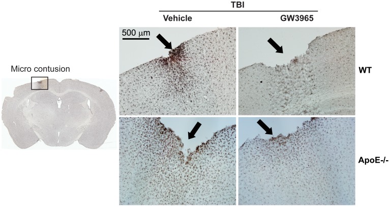
Figure 8. Pronounced microglial activation is localized only around contused areas.
In this study, approximately 4–8% of brains subjected to mrTBI showed micro contusions in the absence of gross skull fracture. The left panel shows a Iba1-stained coronal section at approximately −1.82 mm from bregma [66] with a micro contusion in the cortex below impact site (black square). The right panel shows representative 10X-magnified images of Iba1-stained untreated WT and apoE−/− contused cortices. Microcontusions are denoted by black arrows. Note the pronounced localized activation of microglia around the contusion.

