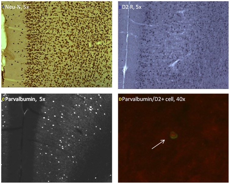
Figure 6. Representative immunohistochemical images of coronal brain sections of the medial prefrontal cortex (mPFC) at postpubertal age.
(A) Neu-N immunolabeling (B) D2 receptor immunolabeling, (C) Parvalbumin immunolabeling and (D) Dual immunolabeling with parvalbumin (Cy3, red) and D2 receptor (FITC, green) specific antibodies.

