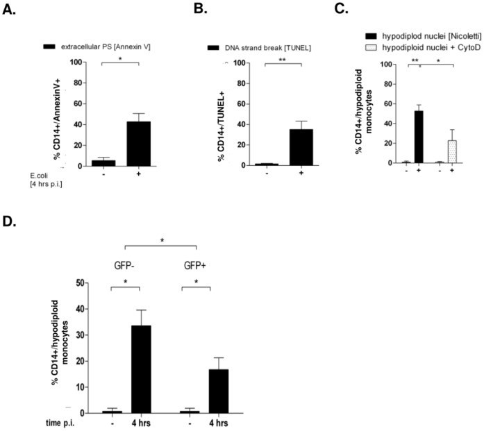
Figure 1. Apoptosis occurs after infection with E. coli-GFP 4 h p.i. in phagocyting and non-phagocyting monocytes.
PBMC were infected with E. coli-GFP for 4 hours and free bacteria were removed. When indicated, CytoD was supplemented 30 minutes prior to infection. Monocyte apoptosis was detected by annexin V (A; n = 5), DNA strand breaks (B; n = 7), and hypodiploidity (Fig. 1C, n = 14, * p<0.05, ** p<0.01). In the same experimental setup, CD14+ monocytes were gated for the absence or presence of GFP-fluorescence and analyzed for their percentage of hypodiploid DNA (Fig. 1D; n = 14, * p<0.05).

