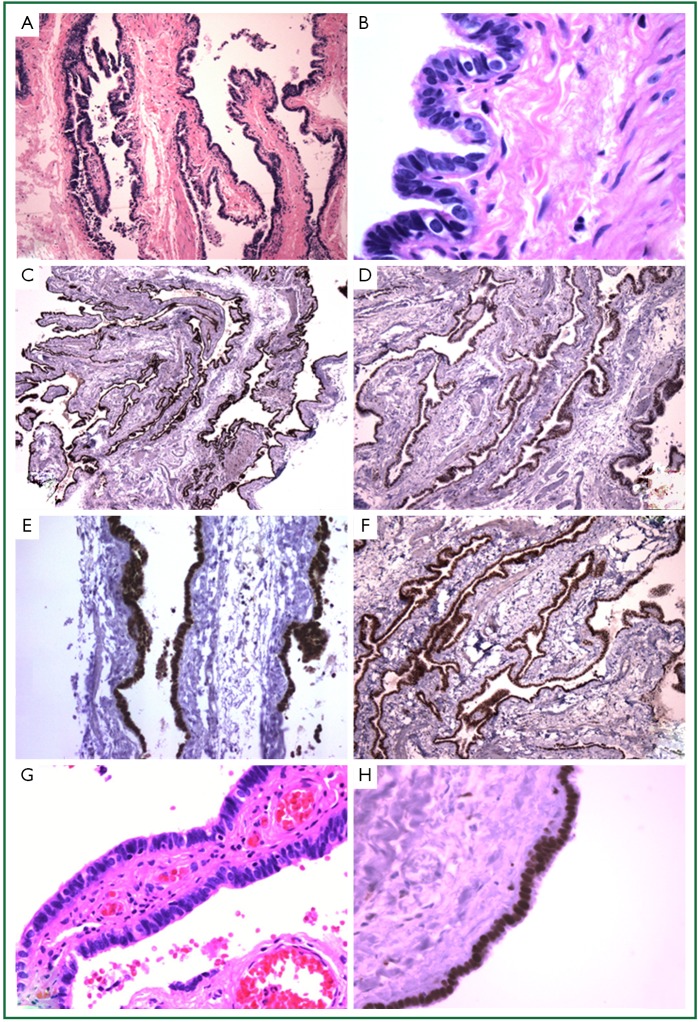
Figure 2.
Histologic appearance of two cases of posterior mediastinal cysts with Mullerian differentiation. (A) Microscopic appearance of cyst from Case 1 showing a thin wall supported by fibrous stroma [hematoxylin and eosin stain (H&E); magnification 4×]; (B) Higher magnification demonstrates the cyst lining is composed of Mullerian-type epithelium with ciliated columnar, secretory, and intercalated cells (H&E, 4×); (C) Immunohistochemistry for ER showing diffuse nuclear positivity in the cyst epithelium (4×); Immunohistochemistry for PR (D), PAX8 (E), and WT-1 (F) also demonstrates diffuse nuclear positivity in the lining epithelium (4×); (G) Microscopic appearance of cyst from Case 2 showing ciliated columnar cells with occasional intercalated cells (H&E, 20×) and also has diffuse nuclear staining for ER (H).

