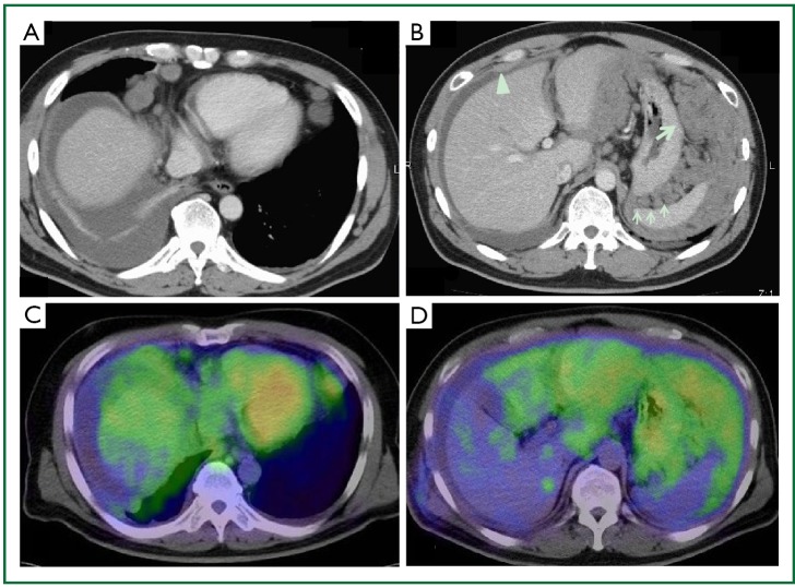
Figure 2.
Chest CT taken on the first visit demonstrates cardiophrenic (A), and intraabdominal lymphadenopathy (B). The CT findings also show a small amount of ascites with smooth parietal peritoneal thickening (B, arrowhead), fine, nodular soft tissue (B, thin arrow), and a large mass-like lesion (B, thick arrow) within the greater omentum, predominantly in the left upper abdomen. PET-CT scan taken 3 weeks after his first visit to our department depicts the intense SUV of subphrenic lymphadenopathy (C), and intraperitoneal cavity (D), especially in the omentum. PET, positron emission tomography.

