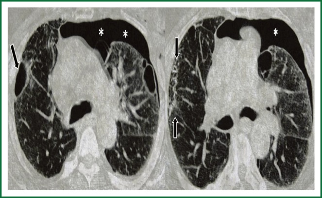
Figure 2.

Axial chest CT demonstrates a pneumothorax in the left [white stars], in the right lobe a cavitary lesion is seen; Axial chest CT at the lower level shows multiple nodular foci [arrows], adjacent to the described cavitary lesion at the right lobe. A pneuumothorax is seen at left [white star].
