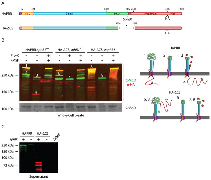
Figure 4.
The region surrounding the native SphB1-dependent cleavage site regulates FHA release and keeps the C-terminus of the prodomain intracellular. (A) Schematic of proteins used in proteinase-K digestion experiments. (B) Top, anti-MCD and anti-HA immunoblot of whole-cell lysate of the cleavage-site deletion strain. Cells were incubated with proteinase-K as indicated. PMSF was added prior to proteinase-K incubation as indicated. Numbers above protein bands correspond to illustrations on the right side of the figure. Illustrations 1–4 represent products of the HAPRR strain, while illustrations 5–9 represent products of the HA-ΔCS strain. These illustrations show how we envision FhaB/FhaC exist at the outer membrane (OM, gray), with the area above the OM representing the extracellular space and the region below the OM representing the intracellular space. Proteinase-K is colored orange. Illustrations are not drawn to scale. Bottom, anti-BvgS immunoblot of whole-cell lysates. (C) Anti-MCD and anti-HA immunoblot of concentrated supernatants. Samples were run on an 8% polyacrylamide gel to resolve the released forms of FHA and prodomain.

