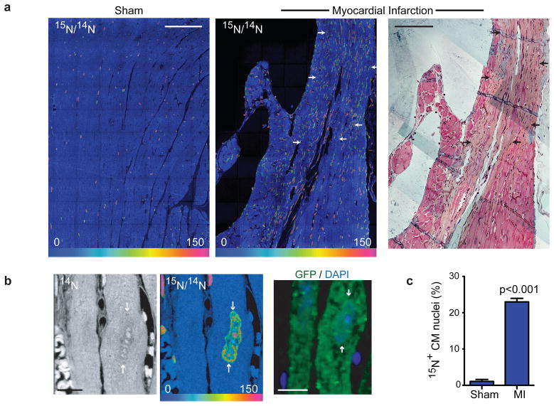
Figure 4. Myocardial injury stimulates division of pre-existing cardiomyocytes.
a) Myocardial infarction (MI) leads to extensive DNA synthesis within and adjacent to scar (arrows). MerCreMer+/ZEG+ mice were treated for 2 wks with 4OH-tamoxifen to induce cardiomyocyte-specific GFP expression before MI or sham surgery, then 15N-thymidine administered continuously for 8wks. Mosaics of 70 60×60μm MIMS tiles. Trichrome stained adjacent section (far right) shows scar. Scale bars = 90μm.
b) 15N-thymidine labeled cardiomyocyte nucleus (white arrows) from MI border region. Immunofluorescent staining demonstrates that the cardiomyocyte is GFP+. Scale bars = 10 μm.
c) Mean % 15N+ cardiomyocyte nuclei after MI (n=4) in the scar border region compared to sham operated mice (n=3). Mean% ± S.E.M.

