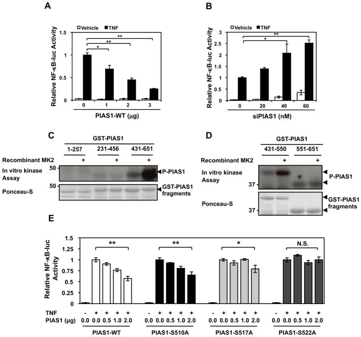
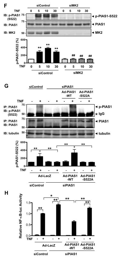
Figure 1. MK2-mediated PIAS1 phosphorylation enhances NF-κB Transactivation under TNF stimulation.
HUVECs were transfected with an NF-κB-luc reporter construct and kinase dead wild-type PIAS1 (A) or PIAS1 siRNA20 (B), stimulated with TNF, and then assayed for firefly and Renilla luciferase activities. For experiments A–B, quantitative data are shown with n=3, values are mean ± S.D.; *P < 0.05, **P < 0.01. (C) PIAS1 was initially divided into three ~200 amino acid fragments (1–257, 231–456, 431–651) and analyzed by in vitro kinase assay. Note strong phosphorylation of the PIAS1 fragment 431–651 by MK2. A representative autoradiogram is shown along with a Ponceau stain to show GST-PIAS1 expression levels. (D) The PIAS1 431–651 fragment was further divided in half (431–550 and 551–651) and analysis was repeated. (E) PIAS1 phosphorylation mutants were constructed and used to evaluate their effects on NF-κB transactivation under TNF stimulation using dual-luciferase reporter assays. *P < 0.05, **P < 0.01 (n=3 mice; mean ± S.D.). (F) HUVECs were transfected for 48 hrs with either MK2 or control siRNA as indicated and then the cells were stimulated with TNF (10 ng/ml) as indicated. PIAS1 S522 phosphorylation was detected by immunoblotting with anti-phospho-PIAS1 S522 (top). The MK2 and PIAS1 expressions were detected by immnoblotting with anti-MK2 and -PIAS1, respectively. Values are mean ± S.D. (lower panel, n=3); **P < 0.01 compared to the sample without TNF stimulation, ##P < 0.01 compared to each concentration of TNF stimulation in the control siRNA. (G) Endogenous PIAS1 in HUVECs was depleted using siRNA, and after 48 hrs the cells were transduced with adenovirus containing PIAS1-WT or PIAS1- S522A mutant. After 16 hrs of transduction cells were stimulated by TNF for 10 min, and PIAS1 was immuno-precipitated by anti-PIAS1 and immunoblotted with anti-phospho-PIAS1 S522 (top). PIAS1 and tubulin expression were detected by immnoblotting with anti-PIAS1 and -tubulin, respectively. Values are mean ± S.D. (lower panel, n=3); *P < 0.05, **P < 0.01. (H) NF-κB transactivation under TNF stimulation was determined using dual-luciferase reporter assays in PIAS1 S522A mutant knock-in experiments. HUVECs were transfected with PIAS1 siRNA or control siRNA and after 48 hrs transduced with Ad-PIAS1-WT, Ad-PIAS1-S522A mutant, or Ad-LacZ. After 16 hrs of transduction the NF-κB-luc reporter was transfected, and cells were stimulated with TNF and then assayed for firefly and Renilla luciferase activities. Values are mean ± S.D. (n=3); *P < 0.05, **P < 0.01.


