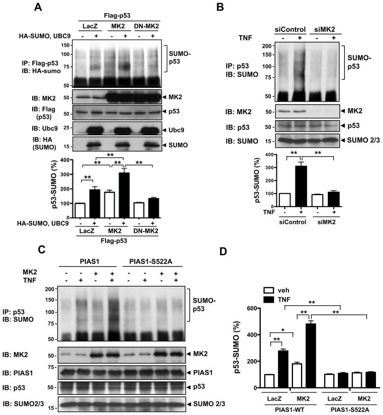
Figure 2. MK2 increases p53-SUMOylation via PIAS1 phosphorylation.
(A) HUVECs were transduced with Ad-LacZ, Ad-MK2, or Ad-DN-MK2 for 18 hrs and then transfected for 24 hrs as indicated with Flag-tagged p53, HA-tagged SUMO3, and Ubc9. p53 SUMOylation was detected by immunoprecipitation with anti-Flag followed by Western blotting with anti-HA (top). Both protein expression and immunoprecipitated p53 were confirmed by anti-Flag and the MK2, Ubc9, and SUMO expressions were detected by indicated antibodies. Mono-SUMOylation band (≈74kDa) and poly-SUMOylation bands (> 78kDa) were detected. (B) HUVECs were transfected for 48 hrs with either MK2 or control siRNA as indicated and then the cells were stimulated with TNF (10 ng/ml) for 3 hrs. p53 SUMOylation was detected by immunoprecipitation with anti-p53 followed by Western blotting with anti-SUMO (top). The MK2, p53, and SUMO expressions were confirmed by immnoblotting with anti-MK2, - p53, and -SUMO, respectively. The quantitative data are shown with n=3 (A and B, lower panel), values are mean ± S.D.; **P < 0.01. (C) HUVECs were transduced for 18 hrs with Ad-PIAS1 or Ad-PIAS1-S522A with Ad-MK2 or Ad-LacZ as indicated, then the cells were stimulated with TNF for 3 hrs. p53 SUMOylation was detected by immunoprecipitation with anti-p53 followed by Western blotting with anti-SUMO (top). PIAS1, MK2, p53, and SUMO expression was confirmed by immnoblotting with anti-PIAS1, MK2, -p53, and -SUMO2/3, respectively. (D) Values are mean ± S.D. (Fig. 2C, n=3); *P < 0.05, **P < 0.01.

