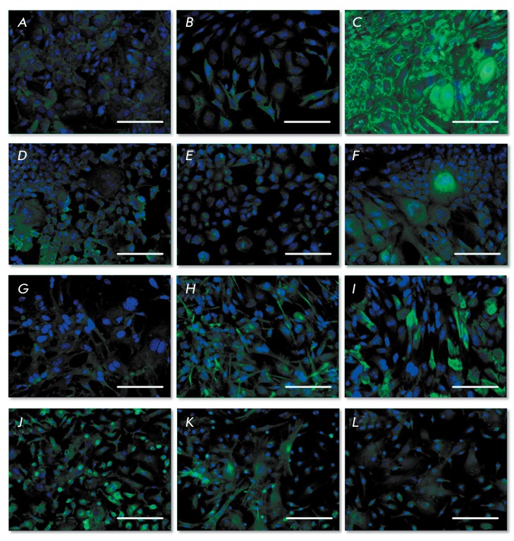
Fig. 3.
Immunocytochemistry of salivary gland and liver progenitor cells on the 1st passage, fluorescent microscopy. Cell nuclei stained by DAPI (blue color), the antigens stained by Alexa Fluor 488 (green color), bar equals to 100 microns. A) PSGC, albumin, 2D conditions; B) PSGC, cytochrome P450 1A1, 2D conditions; C) PSGC, cytokeratin 19, 2D conditions; D) PLPC, albumin, 2D conditions; E) PLPC, cytochrome P450 1A1, 2D conditions; F) PLPC, cytokeratin 19, 2D conditions; G) PSGC, albumin, after cell cultivation for 10 days in the collagen gel; H) PSGC, cytochrome P450 1A1, after cell cultivation for 10 days in the collagen gel; I) PSGC, cytokeratin 19, after cell cultivation for 10 days in the collagen gel; J) PLPC, albumin, after cell cultivation for 10 days in the collagen gel; K) PLPC, cytochrome P450 1A1, after cell cultivation for 10 days in the collagen gel; L) PLPC, cytokeratin 19, after cell cultivation for 10 days in the collagen gel

