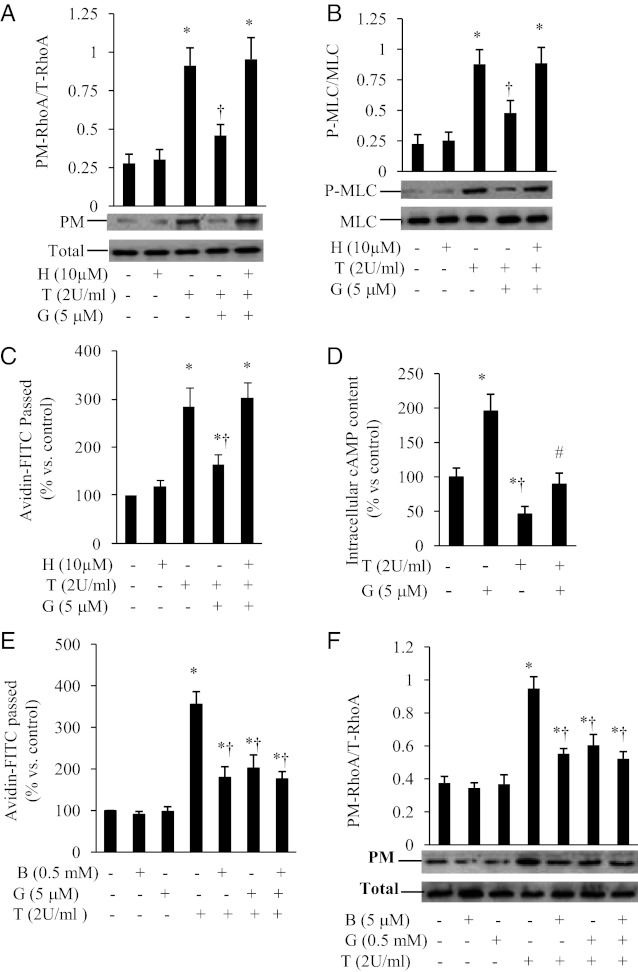
Figure 6.
Genistein inhibition of RhoA and endothelial barrier dysfunction is mediated via PKA. BAECs were preincubated for 30 minutes with 10 μM H89 (H) or vehicle, followed by addition of 5 μM genistein (G) for 30 minutes prior to stimulation with 2 U/ml thrombin (T) for 5 minutes. Cells were then used for measuring RhoA in the plasma membranes (PM) and whole cell extracts (panel A) or for detecting the phosphorylated MLC (P-MLC) and total MLC (panel B). Representative images and quantitative data from four independent experiments are shown. Panel C, BAEC monolayers were preincubated with 10 μM H89 (H) or vehicle for 30 minutes, followed by the addition of 5 μM genistein (G) for 30 minutes prior to stimulation with 2 U/ml thrombin (T) for 1 hour. Avidin-FITC passage was measured. Panel D, BAECs were preincubated with 5 μM genistein (G) or vehicle for 30 minutes followed by the addition of 2 U/ml thrombin (T) for 5 minutes. Intracellular cAMP was measured by EIA. Panel E, BAEC monolayers were preincubated with 0.5 mM 8-bromo-cAMP (B) or 5 μM genistein (G) for 30 minutes. The thrombin-stimulated monolayer permeability was measured. Panel F, BAECs were preincubated with 0.5 mM 8-bromo-cAMP (B) or 5 μM genistein (G) for 30 minutes prior to the addition of thrombin (T; 2 U/ml) for 5 minutes. RhoA in the plasma membranes (PM) and whole cell extracts (Total) were detected by Western blots. Data are expressed as mean ± SE of 3-4 independent experiments, each performed in duplicate. *P < .05 vs control; †P < .05 vs thrombin-alone-treated cells.

