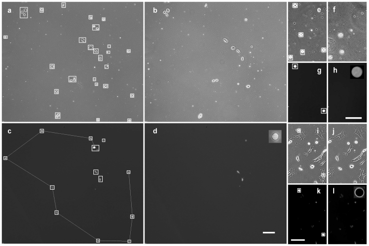
Figure 3. Phase contrast and fluorescent images of cell cultures before and after sorting.
(a,b,c,d) NE-4C, (e, f, g, h) 3T3, ( i, j, k, l) astroglia-microglia cell cultures. (a, c, e, g, i, k) before, (b, d, f, h, j, l) after sorting, i.e., left panels show the cultures before and right panels after sorting. Aperture of the micropipette in action is shown in the insets of (d, h, l). A portion of NE-4C cells and all microglia cells were labeled by GFP. A portion of 3T3 cells were stained by DiI. Sorting process removed these fluorescent cells detected by software. Cells detected in phase contrast and in fluorescent images before sorting are indicated by white frames. Straight lines between cells in (c) show the path of the micropipette: three cells out of the path were excluded from sorting due to a neighbor closer than the sorting resolution of 50 μm, same as the inner diameter of the micropipette. We did not need to detect astroglia cells in phase contrast images (i) because GFP-labeled microglia cells could be removed even from the very close proximity of strongly adherent astroglia cells without perturbing them. Frames in (i) show microglia cells detected in panel k. Scale bars: 100 μm.

