Figure 4.
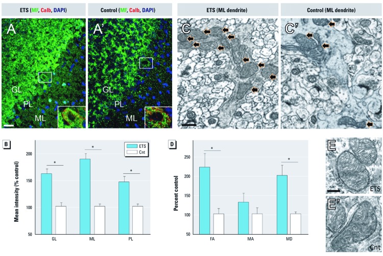
ETS induces Dnm1l-mediated mitochondrial biogenesis, not fragmentation, in cerebellar cortex. (A,A´) The density of mitochondrial staining (MF) increased in ETS300-exposed animals (A) relative to air controls (A´) throughout cerebellar cortex; color-coded representative images; bar = 20 µm; Purkinje cell staining magnified in inset. (B) Mean MF immunofluorescence intensity was significantly greater in granular layer (GL), Purkinje layer (PL), and molecular layer (ML) of cerebellar cortex. (C,C´) Representative electron micrographs show more mitochondria within ML Purkinje dendrites (black arrows) of ETS-exposed animals (C) relative to control (C´). (D) Stereological measures of mitochondrial profiles within the ML; mean mitochondrial fractional area (FA), mitochondrial profile area (MA), and mitochondrial density (MD). (E,E’) Mitochondrial ultrastructure appears healthy and consistent between ETS-exposed (E) and control (E’) animals; representative images from the ML; bar = 200 nm. Data are presented as mean ± SE; n = 4/group. *p < 0.01 compared with controls.

