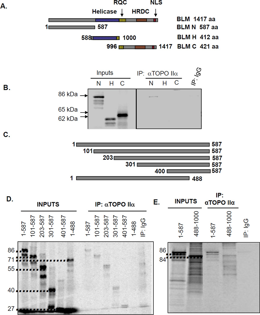
Figure 3. BLM interaction with topoisomerase IIα occurs within the BLM amino-terminus.
(A) Schematic representation of BLM segments tested for topoisomerase IIα binding. RQC identifies the RecQ C-terminal domain, HRDC represents the helicase and Rnase D C-terminal domain, and NLS the nuclear localization sequence. (B) [35S]-methionine-labeled IVTT proteins representing the amino-terminal (N), helicase (H) and carboxy-terminal (C) segments were incubated with topoisomerase IIα and immunoprecipitated (IP) with topoisomerase IIα-specific antibody. Input lanes represent 2% of each IVTT reaction. Control reactions used mαIgG immunoprecipitations. (C) Schematic representation of N-terminal segments. (D) [35S]-methionine-labeled N-terminal segments were generated via IVTT and tested for binding. Input lanes represent 20% of each IVTT reaction. Control reactions used mαIgG immunoprecipitations. (E) [35S]-methionine-labeled IVTT proteins representing the amino-terminal (N) and binding domain tagged to the flanking helicase domain (488-1000) were tested for binding. Input lanes represent 10% of each IVTT reaction. Control reactions used mαIgG immunoprecipitations.

