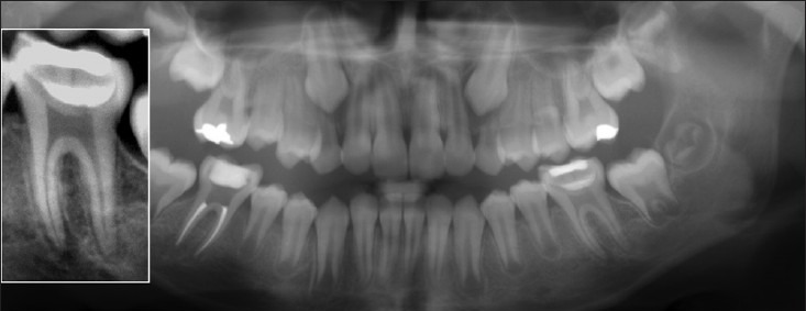
Figure 2.

Post-operative Orthopantomogram (OPG) showing threelayers ofrestorationconsistofindirectpulptherapy/calcium enriched mixture, glass ionomer and composite resin. Left: Higher magnification of treated first lower left molar revealing well defined periapical radiolucency of both roots
