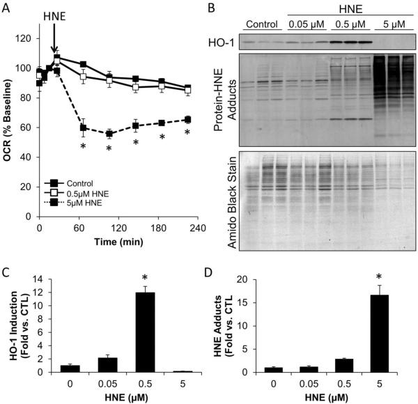
Figure 3. Integration of XF metabolic analysis with western blotting.
The impact of HNE on mitochondrial function, protein modification, and HO-1 induction was examined following OCR measurements in RASMC. Panel A: The OCR of rat aortic smooth muscle cells was measured followed by acute exposure to 0, 0.5, and 5 μM HNE. OCR was then examined for the indicated time. Panel B: At the end of the experiment, cells were lysed in detergent-containing buffer (as described in the text), and the protein was separated by SDS-PAGE, followed by western blotting for protein-HNE adducts and HO-1. The PVDF membrane was then stripped of antibodies and stained with Amido Black (0.1% Naphthol Blue-Black in 40% methanol and 10% acetic acid). The PVDF was then destained with 40% methanol and 10% acetic acid to visualize the separated proteins. Panels C and D: HO-1 expression (Panel C) and protein-HNE adducts (Panel D) were quantified and normalized to Amido Black protein stain. n = 3 per group; *, p≤0.05 vs. cells not treated with HNE.

