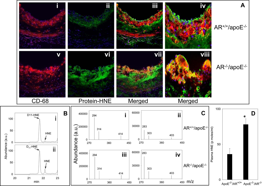
Figure 7.
Genetic ablation of AR increases the accumulation of protein-HNE adducts in the lesions and increases HNE concentration in the plasma. Female AR−/−/apoE−/− and AR+/+/apoE−/− mice (8 weeks old) were maintained on high-fat diet for 12 weeks (Protocol IV). A. Sections of the aortic sinus were stained with Alexa 647 conjugated CD68 (red; i and v) and polyclonal protein-HNE (Alexa 488, green; ii and vi). The yellow fluorescence in the merged image (iii and vii) indicates that protein-HNE co-localizes with macrophages. Nuclei are identified in blue (DAPI). Magnification = 400×. Panels iv and viii show the merged images at 1000× magnification. Plasma HNE was measured by GC-NICI-MS. B. Representative chromatogram of the spectrum of HNE in the plasma of AR+/+/apoE−/− (i) and AR−/−/apoE−/− mice (ii) by select ion monitoring. Panel C shows the spectrum of select ions monitored for the quantification of HNE. D11-HNE was used as an internal standard. D. The group data for plasma HNE levels. * P < 0.01 versus controls.

