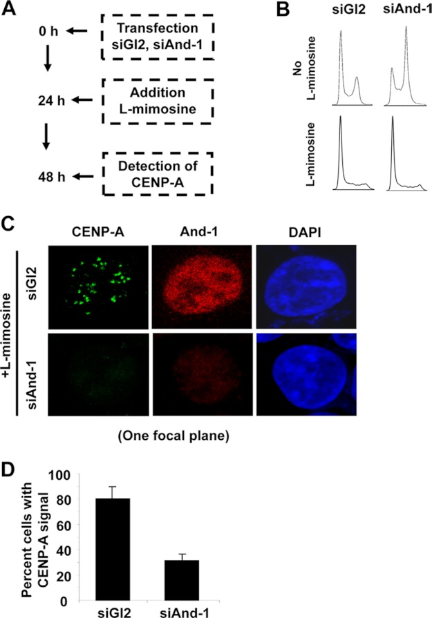
FIGURE 4.
CENP-A localization defects are not due to defects in DNA replication. A, schematic of the experimental procedure. B, FACS analysis of cells harvested as in A. Note the addition of l-mimosine-arrested cells in G1 phase. C, CENP-A centromeric localization reduced in G1 phase cells with down-regulated And-1. CENP-A localization was detected by immunofluorescence (green) in cells harvested as in A. Scale bar, 10 μm. D, quantification of the number of positive cells for centromeric CENP-A signals for HCT116 cells treated as in A. Data represent the means ± S.D. (error bars) from two independent experiments.

