FIGURE 3.

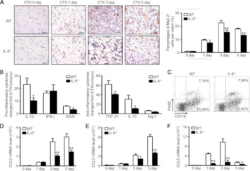
Inflammation response is blunted in IL-6−/− injured muscles compared with WT mice. A, the monocyte/macrophage infiltration in WT and IL-6−/− muscles (0, 1, 3, and 5 days after injury) was examined by immunostaining with anti-Mac-2 (brown) antibody. The right graph indicated the percentages of Mac-2-positive cells per vision (n = 3 in each group). Scale bars, 50 μm. B, at day 3 after injury, proinflammatory and anti-inflammatory cytokines mRNA levels (changed fold compared with uninjured muscle) were accessed by qRT-PCR. C, the percentages of CD11b+ monocytes and CD11b+F4/80+ macrophage in peripheral blood of WT and IL-6−/− mice were detected by FACS. D–F, chemokine CCL2, CCL3, and CCL5 mRNA levels were examined by qRT-PCR at 0, 1, 3, and 5 days after injury (n = 3 in each group). *, p < 0.05; **, p < 0.01 compared with WT values.
