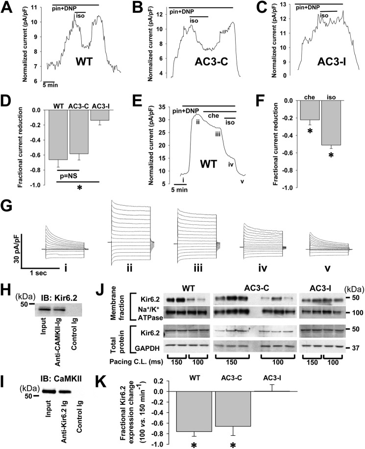
FIGURE 1.
CaMKII effect on native KATP current in isolated ventricular cardiomyocytes. Shown is representative whole cell KATP channel current, stimulated by 100 μmol/liter pinacidil (pin) and 50 μmol/liter DNP, measured during application and wash-out of the CaMKII activator 500 nmol/liter isoproterenol (iso) in isolated cardiomyocytes from WT mice (A), trangenic mice expressing a scrambled, control peptide AC3-C (B), and transgenic mice expressing the CaMKII inhibiting peptide, AC3-I (C). pF, picofarads. D, shown are summary data: AC3-I (n = 12) versus WT (n = 4) and AC3-C (n = 3); *, p < 0.05. E, representative whole cell KATP channel current, stimulated by 100 μmol/liter pinacidil and 50 μmol/liter DNP, was measured during application of the PKC inhibitor, 5 μmol/liter chelerythrine (che), and 500 nmol/liter isoproterenol in an isolated WT murine cardiomyocyte. F, shown are summary data for chelerythrine and isoproterenol effects on pinacidil- and DNP-stimulated KATP channel current in isolated WT murine cardiomyocytes (n = 5; *, p < 0.05 versus base line, the isoproterenol bar represents additional reduction beyond that attributed to chelerythrine application). G, shown is an example of whole cell current tracings measured at points marked from the graph in E. H, On left ventricular lysates from WT mice, immunoprecipitation was performed with the anti-Kir6.2 antibody and probed for CaMKII by Western blot (IB) with the anti-CaMKII Ab. I, immunoprecipitation was performed with the anti-CaMKII Ab and probed for Kir6.2 by Western blot with the anti-Kir6.2 Ab. J, shown are Western blots of biotin-labeled membrane and whole cell fractions of ventricular lysates from isolated hearts paced at 150 or 100 ms cycle length (C.L.). Membrane and total protein fractions are probed with anti-Kir6.2 antibody and with antibodies for Na+/K+ ATPase and GAPDH as controls. K, shown are summary data for the fractional change in Kir6.2 expression with pacing cycle length of 100 versus 150 ms (*, p < 0.05, n = 2 hearts under each pacing condition for WT and AC3-I and n = 3 each for AC3-C).

