
FIGURE 5.
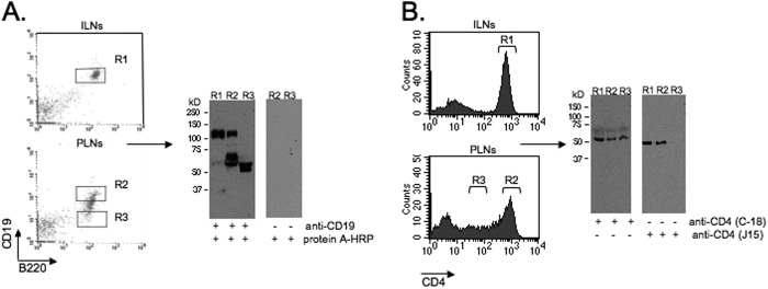
Cleavage of CD4 and CD19 in the PLNs. Shown are the results from Western blot analysis of CD4 and CD19 in cells from inguinal lymph nodes (ILNs) and PLNs, sorted for the high (R1 and R2) and intermediate/low (R3) levels of these markers. The blots were stained with an anti-CD19 antibody that recognizes the cytoplasmic portion of the molecule (A) or two different anti-CD4 antibodies that recognize the intracellular (C-18) or extracellular (J15) portion of CD4 (B). The control blot (A, right panels) was stained with the secondary reagent only. The data are representative of three independent experiments.