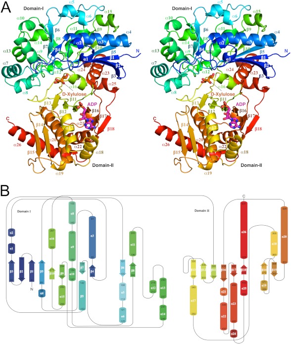
FIGURE 1.
Three-dimensional structure and folding of hXK. A, molecule is shown as a ribbon diagram, in stereo, colored in rainbow style from the N terminus (dark blue) to the C terminus (red). The binding sites for the two substrates are indicated, with d-xylulose shown in orange and the nucleotide (modeled as AMP) shown in magenta, both in stick mode. B, topology diagram, with secondary structural elements colored as in A and labeled.

