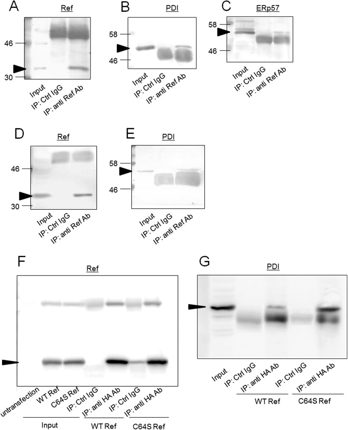
FIGURE 4.
Interaction between PDI and Ref-1. A–E, extracts from GH3 (A–C) or HEK293 (D and E) cells were subjected to immunoprecipitation using anti-Ref-1 antibody. Immunoblotting analyses of precipitated proteins were performed using anti Ref-1 (A), -PDI (B), or -ERp57 (C) antibody (Ab). Arrows indicate signals corresponding to Ref-1, PDI, and ERp57. F and G, HA-tagged wild-type or C64S Ref-1 was expressed in GH3 cells, and immunoprecipitation (IP) was performed using anti-HA antibody or unimmunized mouse serum (control (Ctrl) IgG). Immunoblotting of precipitates was performed using anti-HA (F) and anti-PDI (G) antibody. Arrows indicate signals corresponding to Ref-1 and PDI.

